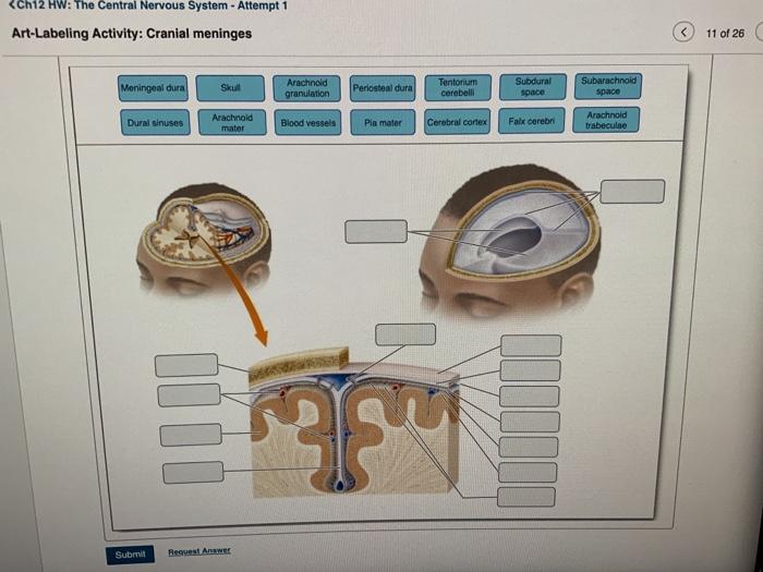
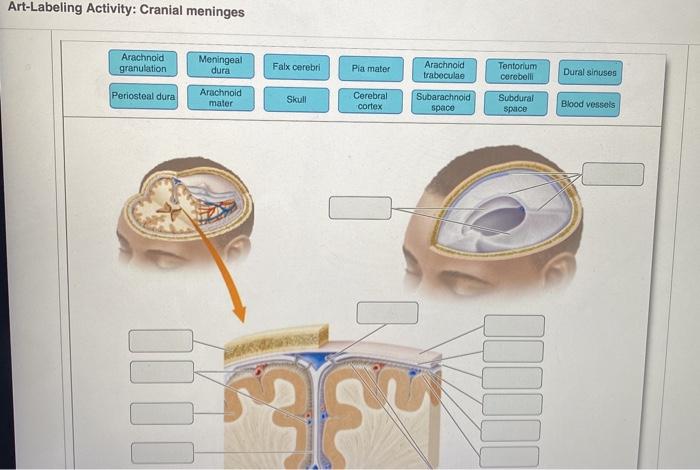
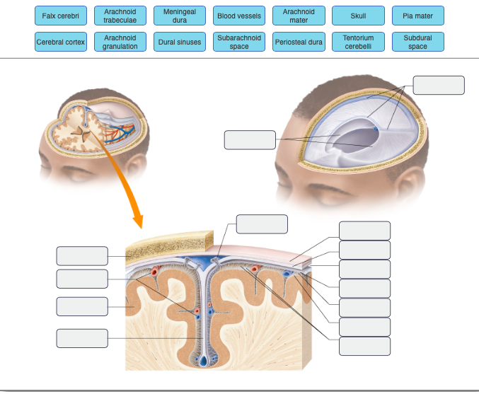
Cranial meninges Arachnold granulation Meningeal dura Falx cerebri Pia mater Arachnoid traboculae Tentorium cerebelli Dural sinuses Periosteal dura Arachnoid mater Skull Cerebral cortex. It connects the brain and spinal cord to the skull and spinal canal.

Labeling Cranial Meninges Overview Diagram Quizlet
Separates the two cerebral hemispheres.

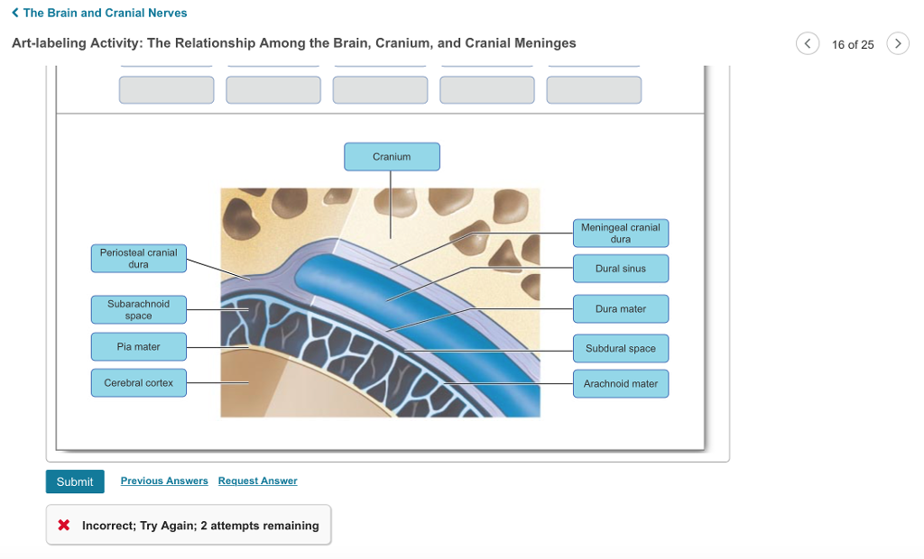
. The Relationship Among the Brain Cranium and. All - ManganatoChapter 5. Brain Cranium and Meninges Close-up View of Cranial Meninges Part A Drag the labels to the appropriate location in the figure.
Up to 24 cash back Bones of the skull Parietal frontal occipital and temporal bones Cranial meninges Dura mater arachnoid mater and pia mater Cerebrospinal fluid Provides protection of the brain and spinal cord Provides support Transports nutrients to the CNS tissue Transports waste away from the CNS Bloodbrain barrier. Pre-Quiz - Part 2. Name and describe the three meninges that cover the brain state their functions and locate the falx cerebri falx cerebelli and tentorium cerebelli.
Which structure is highlighted. Thats why labeling the ear is an effective way to begin your revision. Terms in this set 9 Falx Cerebri.
A set of venous chain that are located between 2 layers of dura mater and drain the cerebral veins of the brain. 122 mV The Brain and Cranial Nerves Art-labeling Activity. Relationships among the brain cranium and meninges Label the major anatomical features of the brain and meninges.
View the full answer. Anatomy of the Heart 79 Exercise 30 Review Sheet Art-labeling Activity 1 1 of 2 Identify the structures of the heart. Which of the following is the outer layer of the meninges.
34 transverse cervical nerve. Figure 251 2 of 3 Art-labeling Activity. Thorpe Park Early Entry Art Labeling Activity Cranial Meninges Quizlet Grams Express Lewisburg Menu Amber Cove Dolphin Swim Treetops Resort Gaylord Mi Screaming Christmas Goat Toy Tractor Supply Pantheon Guide Support Importance Of Classroom Participation Pdf American Airlines At Smith Reynolds Airport SitemapSitemap.
Exam 1 Review - Lecture notes. The meninges forms a protective barrier that safeguards the sensitive organs of the CNS against trauma. What organ system controls the activity of the eccrine sweat glands.
100 7 ratings The three layers of membranes are called as the meninges and. Figure 142a The Spinal Cord and Spinal Meninges Anterior view of spinal cord showing meninges and spinal nerves. Digestive System - Medical Terms.
Compare and contrast the meninges of the spinal cord and the brain. Cranial meninges Part A Drag the appropriate labels to their respective targets. Seeing them all together in this way is a great way to learn since anatomical structures do not exist in isolation.
œ It is the soft tissuess that covers the vault of skull. The skull the ver-tebral column and the thoracic cage. Try to understand and memorize what you can from the labeled diagram then try to label the cranial nerves yourself with our cranial nerves labeling quiz exercise available to download below.
However review of the embryology and anatomy reveals the dura to be a complex vascularized and innervated structure not a simple fibrous covering. œ It extends from the superciliary arches eye brows anteriorly to the superior. Make use of your textbook and atlas during this time.
This is a great way to start to get the cogs turning and warm up your memory before you take our other cranial nerve quizzes but one. This shows you all of the structures youve just learned about in the video labeled on one diagram. Free labeling quiz.
It also contains an ample supply of blood vessels that deliver blood to CNS tissue. Anatomy and Physiology Art-Labeling Activity. 8192015 3 BASIC STRUCTURE OF THE BRAIN AND SPINAL CORD Spinal cord long tubular organ enclosed within protective vertebral cavity.
For this view the dura and arachnoid membranes have been cut longitudinally and retracted pulled aside. 36 superior root of Ansa cervicalis nerve. Recovery of 14 Clactate and other labeled compounds in the meninges after microinfusion of 14 C-labeled glucose or lactate into the inferior colliculus with 34 of the glucose-derived label recovered in the meninges compared with 60 in the infused inferior colliculus brought our attention to lactate discharge via perivascular-lymphatic.
Ends between first and second lumbar vertebrae 43 46 cm 17 18 inches in length and only ranges from 065125 cm 02505 inches in diameter Central canal an internal cavity within. The meninges functions primarily to protect and support the central nervous system CNS. Meninges are connective tissue membranes that line the neurocranium and vertebral canal and enclose the central nervous system cns brain and spinal cord.
61 mV C- 122 D244 mV E. Identify the cranial nerves by number and name on a model or image stating the origin and function of each. Part A Drag the labels to the appropriate location in the figure.
In its mainstream origins it was more common for a fighter to only be trained in one or two martial arts but nowadays it is far more likely that a contender must be versed in a minimum of. See more ideas about taekwondo tattoo taekwondo martial arts. 33 great auricular nerve.
Which cranial nerve carries visual information from the eyeball to the brain. 32 lesser occipital nerve. Circulation of Cerebrospinal Fluid Answer the following questions.
ErosionAUTOMOTIVE ELECTRICAL AND ENGINE PERFORMANCEart labeling activity cranial meninges - academicaaseroManga List - Genres. Cranial meninges Part A Drag the appropriate labels to their respective targets. Notice the blood vessels that run in the subarachnoid space bound to the outer surface of the delicate pia mater.
Discuss the formation circulation and drainage of cerebrospinal fluid. See more ideas about art tattoo martial arts tattoos tattoo designs. Like the dorsal cavity the ventral cavity has two subdivisions.
37 inferior root of Ansa cervicalis nerve. Of the three layers of the meninges the dura mater the. An Introduction to Brain Structures 10125 Left cerebral Midbrain Fissure Medulla oblongata Pons Cerebellum Cerebrum Spinal cord Sulci Brai The Brain and Cranial Nerves Art-labeling Activity.
Up to 10 cash back The dura is traditionally viewed as a supportive fibrous covering of the brain containing the dural venous sinuses but otherwise devoid of vessels and lacking any specific function. Take a moment to look at the ear model labeled above. Brain Cranium and Meninges Close-up View of Cranial Meninges Part A Drag the labels to the appropriate location in the figure.
Secretes CSF allows CSF to be reabsorbed into blood acts as the bloodbrain barrier filters CSF. Blends with inferior portion of brainstem.
Solved Ch12 Hw The Central Nervous System Attempt 1 Chegg Com

A P 1 Chapter 13 Mastering Assignments Flashcards Quizlet
Solved The Brain And Cranial Nerves Art Labeling Activity Chegg Com

Pin On Radiology

Anatomy 2220 Brain And Sp Lab Review Flashcards Quizlet
Solved Art Labeling Activity Cranial Meninges Arachnold Chegg Com
The Brain Cns Lecture 12a Biol Ppt Video Online Download
Solved Meningealblood Vessels Arachnoid Mater Arachnoid Chegg Com
0 comments
Post a Comment